Ultrasound Used for BPH Diagnosis
What is an Ultrasound?
Ultrasound for an enlarged prostate is a diagnostic imaging technique used to assess the prostate gland's size, shape, and condition. It involves using sound waves to create images of the prostate gland and the surrounding tissues.
This procedure helps doctors evaluate the size of the prostate gland and detect any abnormalities or growths, which can be indicative of conditions like benign prostatic hyperplasia (BPH) or prostate cancer.
Purpose of Ultrasound
- Assessment of Prostate Size
- Detection of Abnormalities
- Assessment of bladder and emptying
- Monitoring disease or treatment progression
Who is Suitable for Ultrasound?
Suitable candidates for ultrasound evaluation may include:
- Men with Lower Urinary Tract Symptoms (LUTS): Individuals experiencing symptoms such as frequent urination, urinary urgency, difficulty initiating or maintaining urination, weak urine stream, or incomplete bladder emptying may benefit from ultrasound assessment of the prostate gland to determine if BPH or other prostate-related conditions are contributing to their symptoms.
- Men with Elevated Prostate-Specific Antigen (PSA) Levels: Ultrasound imaging may be recommended for men with elevated PSA levels to assess the prostate gland's size and condition and evaluate for any abnormalities requiring further investigation.
- Men with a Family History of Prostate Conditions: Individuals with a family history of prostate conditions, such as BPH or prostate cancer, may have an increased risk of developing these conditions themselves. As such, doctors may recommend ultrasound imaging as part of routine screening or diagnostic evaluation for men with a familial predisposition to prostate conditions.
- Men with Prior Prostate Conditions: Individuals with a history of prostate conditions, such as BPH or prostate cancer, may require periodic ultrasound monitoring to assess disease progression, treatment response, and overall prostate health. Ultrasound imaging can provide valuable insights into changes in the size and structure of the prostate gland over time, helping doctors optimise management strategies accordingly.
Benefits of Ultrasound for BPH
Ultrasound imaging offers several benefits in diagnosing and managing prostate conditions. Here are some of the key advantages:
- Non-Invasive: Ultrasound imaging is a non-invasive procedure that does not require ionising radiation or surgical incisions. It involves using sound waves to create detailed images of the prostate gland and surrounding tissues, making it a safe and well-tolerated diagnostic option for individuals with prostate-related concerns.
- Real-Time Imaging: Ultrasound provides real-time images of the prostate gland, allowing doctors to visualise the structure, size, and condition of the prostate in motion. This real-time feedback can be invaluable for assessing dynamic processes within the prostate, such as changes in urine flow or abnormalities during biopsy procedures.
- Readily available: Compared to other imaging modalities, such as magnetic resonance imaging (MRI) or computed tomography (CT) scans, ultrasound imaging is often more available and cost-effective. It requires less expensive equipment and can be performed quickly in a clinical setting. It is a practical option for routine screening, diagnostic evaluation, and follow-up monitoring of prostate conditions.
- No Radiation Exposure: Unlike imaging techniques such as CT scans or X-rays, ultrasound imaging does not expose patients to ionising radiation. This is particularly advantageous for individuals who require repeated imaging studies over time, as it minimises the risk of cumulative radiation exposure and associated health concerns.
- Versatility: Ultrasound imaging can be used for various purposes in evaluating prostate conditions, including assessing prostate size, detecting abnormalities, guiding biopsy procedures, and monitoring disease progression.
Types of Ultrasound
Several types of ultrasound imaging techniques may be utilised for evaluating an enlarged prostate. These include:
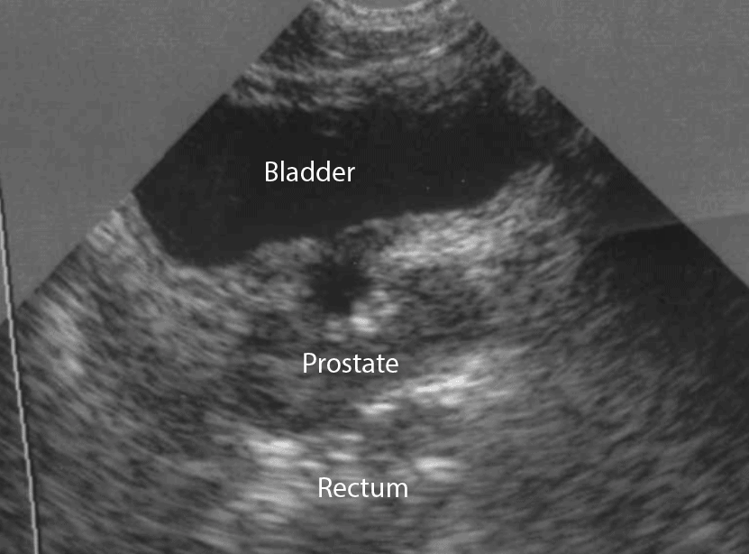
- Transrectal Ultrasound (TRUS) is the most common imaging used to assess the prostate gland. During a TRUS procedure, a small transducer probe is inserted into the rectum to obtain images of the prostate gland and surrounding tissues. This approach provides detailed pictures of the prostate and allows for an accurate assessment of prostate size, shape, and abnormalities.
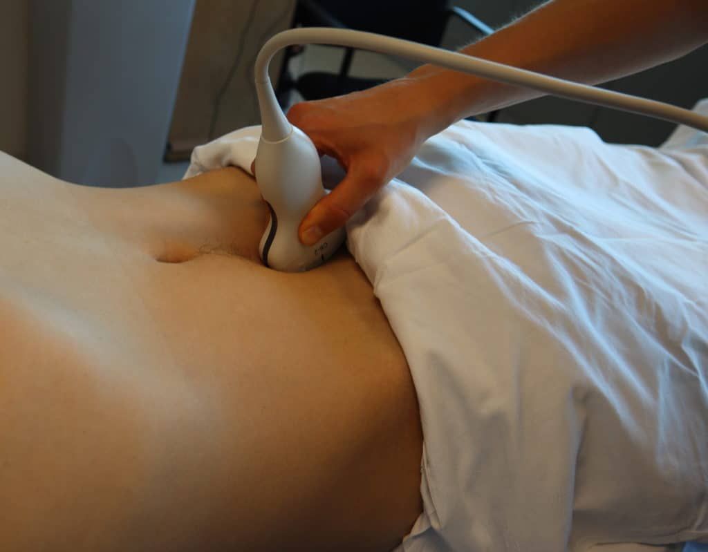
- Transabdominal Ultrasound involves placing the ultrasound transducer on the abdomen, over the lower part of the abdomen, to visualise the prostate gland. While transabdominal ultrasound can provide some information about the prostate, it may offer a different level of detail than transrectal ultrasound, particularly for evaluating the internal structures of the prostate gland.
- Three-dimensional (3D) Ultrasound imaging uses advanced technology to create three-dimensional images of the prostate gland. This technique allows the doctor to visualise the prostate from multiple angles and perspectives, enhancing their ability to assess the prostate's size, shape, and abnormalities with greater accuracy.
- Doppler Ultrasound is a specialised technique that assesses blood flow within the prostate gland and surrounding tissues. Doppler ultrasound can help identify areas of increased vascularity by measuring blood flow patterns and velocities.
- Contrast-enhanced ultrasound (CEUS) involves using contrast agents, such as microbubbles, to improve the visualisation of blood flow and tissue perfusion within the prostate gland. This technique can provide additional information about vascularity and tissue characteristics, aiding in detecting and characterising prostate abnormalities.
Preparation Before an Ultrasound
Before an ultrasound, there are several steps you can take to ensure a smooth and successful procedure:
- Follow Preparatory Instructions: You must often be well-hydrated with a full bladder before presenting for ultrasound. The clinic conducting the test will advise you on how much fluid to drink and when.
- Empty Your Bladder:
During the ultrasound examination, you may be asked to empty your bladder. This can help determine the residual volume, indicating potential urinary obstruction or bladder dysfunction. the
- Bring a List of Medications: It can be helpful to bring a list of medications you currently take to your ultrasound appointment.
- Wear Comfortable Clothing: For your ultrasound appointment, choose comfortable, loose-fitting clothing. You may be asked to change into a gown or remove certain clothing items during the procedure.
Ultrasound Procedure
During an ultrasound for an enlarged prostate, the following steps typically occur:
- Preparation: You may be asked to change into a gown and lie on an examination table, either on your side with your knees bent or in a different position that allows access to the rectum for transrectal ultrasound imaging.
- Transducer Insertion: A lubricated transducer probe will be placed on your lower abdomen. The transducer emits sound waves that bounce off the prostate gland and bladder, creating detailed images displayed on a monitor.
- Image Acquisition: The doctor will move the transducer probe back and forth to obtain images of the prostate gland and surrounding structures. You may be asked to hold still or change positions slightly to facilitate imaging of different areas.
- Evaluation: As the images are obtained, the sonographer will evaluate the size, shape, and condition of the prostate gland, as well as any abnormalities or areas of concern that may be present.
What to Expect After an Ultrasound?
- Resume Normal Activities: You can resume your normal activities immediately after the ultrasound procedure. There are typically no restrictions on driving, returning to work, or engaging in other daily activities.
- Discuss Results: Your doctor will review the ultrasound images and discuss the findings with you. Depending on the results, further evaluation or treatment options may be recommended.
- Follow-Up: Depending on the reason for the ultrasound and the examination findings, your doctor may recommend follow-up appointments or additional tests to evaluate any abnormalities further or monitor your condition over time.
- Report Any Concerns:
If you experience any unusual symptoms or have concerns after the ultrasound procedure, such as persistent pain, bleeding, or signs of infection, contact your doctor for further evaluation and guidance.


